

Simulation Cases Cliff Notes April 2017
/
Every month we summarize our simulation cases. No deep dive here, just the top 5 takeaways from each case (blast injury, electrical injuries, pelvic fractures, pediatric burns).
Blast Injuries

https://commons.wikimedia.org/wiki/File:Blast-wave.png
1. Blast injuries comprises a multitude of different potential injuries secondary to an explosion.
An explosion is an event that occurs when a substance rapidly releases energy and produces a large volume of gaseous products.
The extreme compression of molecules by this change in energy creates the blast wave that moves outward from the epicenter of the blast.
Injuries seen from an explosion are dependent upon the size of the explosive charge, the nature of the explosive, the container, any shielding or protective barriers between the victim and the explosion, the surrounding environment, the method of delivery, and the distance between the explosion and the victim.
2. The first priority of the emergency physician who is faced with the aftermath of an explosion is to activate the hospital’s external disaster plan.
Are you aware of your hospital’s disaster plan? Make sure your hospital has one and you know how to activate it.
While complex and involving multiple players, key aspects include:
Clear the ED of all possible patients by either discharge or admissions before the first patients of the explosion arrive.
Hospital administration should simultaneously cancel all elective surgery cases, clear the recovery room, and clear as many intensive care beds as possible.
3. Trauma from explosions have traditionally has been categorized according to the following scheme and can help the emergency provider anticipate potential injuries.
PRIMARY INJURY- Caused by direct effect of blast wave
SECONDARY INJURY- Caused by other objects that are accelerated by the explosive wave (i.e. flying glass)
TERTIARY INJURY - Effects caused by movement of victim and being thrown against other objects (i.e. thrown against wall)
QUATERNARY INJURY - Burns from fire or radiation, crush injury associated with structural collapse, poisoning from carbon monoxide or other toxic products of the explosion, and inhalation of dust or chemicals from the explosion

http://www.tamingthesru.com/blog/grand-rounds/recap-52015-disaster-day
4. The air/lung interfaces are very susceptible to a blast wave. Lung injury is the most common fatal injury from an explosion. Blast lung (BL) is the most common fatal injury caused by a primary blast among initial survivors[1].

httpblog.clinicalmonster.com201609listen-bass-go-boom
Pulmonary barotrauma from a pressure wave includes pneumothorax, pneumomediastinum, pulmonary contusion/hemorrhage, and air embolism form bronchopleural fistulas.
BL is characterized by a triad of dyspnea, bradycardia, and hypotension. Other common findings include wheezing, cough, hemoptysis.
It is the most common fatal injury caused by the primary blast among the initial survivors of the explosion.
A simple frontal chest x-ray is diagnostic for most cases of pulmonary barotrauma from blast. Blast lung produces a characteristic “butterfly” pattern on chest x-ray [2].
5. Explosions have the potential to cause both blunt and penetrating multi-system injuries. Care of these trauma patients and these injuries should follow typical ATLS protocols. A few key differences include:

OUr POOR Mannequin Takes such a beating for our residents!
A blast wave compresses tissues and the resulting forces exceed the tensile strength of the material and causes shearing of vascular beds, pulmonary contusions, and gastrointestinal hemorrhages as the tissues are compressed and expanded.
Blast lung should be anticipated.
Gastrointestinal injury (mesenteric bleeding, bowel contusion, colonic perforation) from a blast wave is inconsistent in presentation and may not be apparent externally.
Secondary blast injury (much more common than primary blast injury) caused by bomb fragments and other debris propelled by the explosion can have very minor appearing external wounds. Use radiographs judiciously to find foreign bodies.
Tertiary blast injuries and deceleration by impact into a rigid structure can cause any type of blunt trauma.
Miscellaneous quaternary injuries could include burns from fire or radiation, crush injury associated with structural collapse, poisoning from carbon monoxide or other toxic products of the explosion, and inhalation of dust or chemicals from the explosion.
References
1. Yelverton J. Blast biology. In: Cooper C, Dudley H, Gann D, eds. Scientific Foundations of Trauma. 1st ed. Oxford, UK:
Butterworth-Heinemann; 1997:189-199. (Textbook chapter)
2. Argyros GJ. Management of primary blast injury. Toxicology 1997;121(1):105-115. (Review)
Electrical Injuries
1. The damage incurred during an electrical injury depends upon the voltage, the resistance of tissues, the amperage (or current strength), the type of circuit (direct or alternating current), the current pathway, and the duration of contact.
Plumbing analogy:

http://www.creativeoutdoorlighting.com/electrical-plumbing-analogy/
Amperage is the volume of water.
Resistance is the diameter of pipe (bone, tendon and fat have highest resistance and typically worse damage).
Voltage is the difference between entrance and exit pressure of pipe.
2. Electrical injuries are traditionally divided into high-voltage vs low-voltage exposures, AC vs DC circuits. Understanding these distinctions can help you anticipate potential injuries.

http://www.abb.com/cawp/seitp202/5e61baec6e0361f4c12579dd0040522f.aspx
AC vs DC
AC
Electrons flow back and forth through a conductor in a cyclical fashion.
Found in most offices and and homes.
Causes tetany that prolongs contact with source, making it potentially more dangerous.
It has been suggested that ventricular fibrillation is more common with low-voltage AC injuries as it increases likelihood of current going through the heart during vulnerable relative refractory period [1].
DC
Used in batteries, automobile electrical systems, high voltage power lines.
Causes a single muscle contraction that throws victims from electrical sources.
Asystole is seen more often with DC high-voltage injuries[2].
High voltage (>1000 volts)
Direct contact with body (can cause massive underlying damage to soft tissue and bone that may not be apparent from the surface).
Flash injuries (caused by arc between source and patient; can generate temps up to 5000C (9032F) and ignite clothing).
Low-voltage (<1000 volts)
Common household circuits in the United States and Canada, which provide 120 volts for general use and 240 volts for high-power appliances.
Tend to cause small, well demarcated contact burns at entrance and exit site.
3. Electrical injuries have three main mechanisms of injury.
Direct trauma from electric current coursing through body
External trauma still may not represent.
It has been suggested that ventricular fibrillation is more common with low-voltage AC injuries, whereas asystole is seen more often with DC high-voltage injuries [1].
Overall, exposure to low-voltage AC is most likely to cause cardiac consequences as it increases the likelihood of current flow through the heart during the vulnerable relative refractory period.
Trauma from conversion of electrical energy to thermal injury
High-voltage injuries may largely spare the skin surface but cause massive damage to underlying soft tissue and bone, necessitating escharotomies, fasciotomies, or amputations [2,3].
Damage to intima of vessels may result in thrombosis and occlusion immediately or over several days.
Cardiac damage is possible from the transcardiac passage of electrical current.
Damage to intima of vessels may result in thrombosis and occlusion immediately or over several days.
Incidence of spinal cord injury following high-voltage electrical trauma ranges from 2-27% [3].
Cataracts and hearing loss have also been described.
Mechanical effects of the electric current include violent contractions and falls
Exposure to DC causes a single muscle contraction that throws the victim away from the electrical source.
AC causes tetany that prolongs contact with the source, making it potentially more dangerous.
4. Delayed arrhythmias are rare and tend to occur only in patients with an arrhythmia on presentation, as has been shown in multiple prospective and retrospective trials encompassing both low-voltage and high-voltage injuries [4-10].

https://lifeinthefastlane.com/ecg-library/atrial-fibrillation/
Obtain an initial ECG in the ED on all patients with an electrical injury, regardless of the voltage.
Traditionally, low-voltage electrical exposure who have a normal ECG on presentation and no loss of consciousness or arrhythmia can be discharged without cardiac monitoring.
Patients with history of loss of consciousness, documented arrhythmias either before or after arrival to the ED, ECG evidence of ischemia, or who have sustained a high voltage electrical injury should be admitted for additional monitoring.
5. All electrical injuries should be evaluated and managed as multi-system injuries along with the following unique considerations:
All patients should receive an ECG and be placed on cardiac monitor to screen for cardiac injury and arrhythmias.
Urinalysis and CK levels should be obtained with high voltage injuries to assess muscle injury/rhabdomyolysis.
Extremity burns should be followed closely for possible fasciotomy (progressive neurologic dysfunction, vascular compromise, increased compartment pressures, vascular compromise).
The fluid requirements of patients with electrical injuries are generally much greater than those of patients with thermal burns. Because cutaneous burns from electrical injuries do not represent the full extent of injury, formulas for fluid resuscitation based on percentage of body surface area burned are not applicable. Fluid resuscitation should maintain a urine output of 1.0 to 1.5 cc/kg per hour.
References
1. Lown B, Neuman J, Amarasingham R, Berkovits BV. Comparison of alternating current with direct electroshock across
the closed chest. Am J Cardiol. 1962;10:223-233.
2. Arnoldo BD, Purdue GF, Kowalske K, Helm PA, Burris A, Hunt JL. Electrical injuries: a 20-year review. J Burn Care &
Rehabil. 2004;25(6):479-484.
3. Varghese G, Mani MM, Redford JB. Spinal cord injuries following electrical accidents. Paraplegia. 1986;24(3):159-166.
4. Rai J, Jeschke MG, Barrow RE, Herndon DN. Electrical injuries: a 30-year review. J Trauma. 1999;46(5):933-936.
5. Blackwell N, Hayllar J. A three year prospective audit of 212 presentations to the emergency department after electrical
injury with a management protocol. Postgrad Med J. 2002;78(919);283-285.
6. Bailey B, Gaudreault P, Thivierge RL. Cardiac monitoring of high-risk patients after an electrical injury: a prospective
multicentre study. Emerg Med J. 2007;24(5):348-352.
7. Bailey B, Gaudreault P, Thivierge RL, Turgeon JP. Cardiac monitoring of children with household electrical injuries.
Ann Emerg Med. 1995;25(5):612-617.
8. Bailey B, Gaudreault P, Thivierge RL. Experience with guidelines for cardiac monitoring after electrical injury in
children. Am J Emerg Med. 2000;18:671-675.
9. Garcia CT, Smith GA, Cohen DM, Fernandez K. Electrical injuries in a pediatric emergency department. Ann Emerg
Med. 1995;26(5):604-608.
10. Purdue GF, Hunt JL. Electrocardiographic monitoring after electrical injury: necessity or luxury? J Trauma.
1986;26(2):166-167.
PELVIC FRACTURES

https://commons.wikimedia.org/wiki/File:Diastasis_symphysis_pubis_1300500.JPG
1. Assess the mechanism of the pelvic injury (lateral compression, AP compression, vertical shear) and determine the fracture pattern.
Vertical shear and AP compression tend to be the highest risk for bleeding and hemodynamic instability
“Open Book” pelvic fractures requires immediate external fixation (pelvic binder).
2. Open book pelvic fracture is a life threat – a “Circulation” issue/life threat in our primary survey. Assess for unstable pelvic fracture in your primary survey and address it here with a pelvic binder.
Video credit to www.tamingthesru.com
3. Bony stability does not equal hemodynamic stability.
4. Initial management should include application of a pelvic binder, crystalloids, and early blood. If the patient is unresponsive to these first tier interventions, mobilize the next tier of treatment (angiographic embolization). Consider initializing the massive transfusion protocol.
5. Pelvic trauma is a multidisciplinary sport (think of all the consultants that might be needed to help manage pelvic injuries (i.e. trauma surgeon, urologist, orthopedist, IR). Mobilize help early.
Pediatric Burns
1. Burns in patients < 14 yo are consistently among the top causes of injury induced mortality (#3 after motor vehicle accidents and accidental drowning). Scalds (from hot liquids) comprise the majority of pediatric burns[1].
2. Initial management of the burn patient should focus on:
Airway assessment
Evaluation for signs of inhalational injury
Evaluation for concomitant trauma
Prevention of burn process (remove all affected clothing, cooling with cool water/saline).
Removing rings/constrictive jewelry and clothes
3. Your history and physical exam should focus on the type of burn, burn depth, burn severity and % Total Body Surface Area (TBSA).

http://www.burn-recovery.org/injuries.htm
What type of burn was it (scald, flame, contact, electrical, friction, sun)?
Burn classification
Depth as established by American Burn Association
Superficial - epidermis only
Superficial partial thickness - epidermis and extends down to papillary upper dermis; wet and weeping, often with blisters
Deep partial thickness - dry and white (easy way to differentiate between them is the ability to blanch)
Full thickness - extends through dermis and into underlying adipose tissue
Severity as established by American College of Surgeons
Minor: < 10% TBSA or full thickness involving < 2% TBSA
Moderate: 10-20% TBSA of partial thickness but < 10% full thickness TBSA
Severe: > 20% TBSA partial thickness, > 10% full thickness burns
Percentage TBSA burn using Lund-Browder chart
More accurate than rule of 9’s [2]
Only partial and full thickness burns count toward %TBSA
TBSA determined based on this TBSA – over and under fluid resuscitation has own complications

4. Evaluate for and treat concomitant trauma associated with burns.

https://lifeinthefastlane.com/unusual-urine-004/
Electrical burn?
Consider myoglobinuria, edema, compartment syndrome, cardiac dysrhythmias
Structure fire?
Consider trauma from collapsing structure, carbon monoxide and cyanide poisoning
Hydrofluoric acid burn?
Consider hypocalcemia
5. The primary pillars of burn treatment includes airway management, fluid resuscitation, wound care, and pain management.
https://www.hospira.com/en/products_and_services/iv_solutions/LACTATED_RINGERS_INJECTION
Airway management
Eval for stridor, hoarseness, sob, singed nasal hairs, cough, soot in oral cavity, history of being in fire in enclosed space
Use largest tube possible to improve bronchoscopy later
Burn shock and fluid resuscitation
Only for burns > 20% TBSA [3]
Use either Galveston or Parkland formula
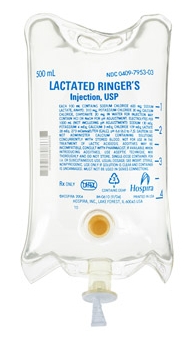
Use Lactated Ringers (sodium content (130 meq/L) capable of correcting hyponatremia caused by burn [3]
Parkland formula: (4 mL/kg x %TBSA) + normal 24 hour maintenance
½ given in first 8 hours after onset of burn
Remainder given in remaining 16 hours
These formulas are a general guide for where to start fluids
Monitor ongoing fluid requirements with UOP (1-2 ml/kg/h), input/output hemodynamics should be used to tailor fluid requirements to patient needs

http://www.airlats.com/egg-shells-hold-key-to-rapid-healing/
Wound care
Silver sulfadiazine commonly available, but no evidence to show more effective than petroleum jelly products [4,5]
Sterile saline and gauze to gently remove sloughing epidermis [6]
General consensus regarding blister management:
Blister intact and < 1 cm2, leave intact.
If large or rupture imminent, may be aspirated and debrided
Blisters > 1 cm2 can be debrided or left intact; there is contradictory evidence on topic
Once burn cleaned, topical antimicrobial agent (SS, Mupirocin, polysporin, bacitracin) can be applied
Consider amnion membrane and membranous dressings (eg. Biobrane®, DuoDERM®)
They don’t have to be changed, they promote faster healing, improve pain relief [7]
Burns are painful. Administer pain medication liberally.
References
1. Shields BJ, Comstock RD, Fernandez SA, et al. Healthcare resource utilization and epidemiology of pediatric burn-associated hospitalizations, United States, 2000. J Burn Care Res. 2007;28(6):811-826.
2. Wachtel TL, Berry CC, Wachtel EE, et al. The inter-rater reliability of estimating the size of burns from various burn
area chart drawings. Burns. 2000;26(2):156-170.
3. Pham TN, Cancio LC, Gibran NS. American Burn Association practice guidelines burn shock resuscitation. J Burn Care
Res. 2008;29(1):257-266.
4. Genuino GA, Baluyut-Angeles KV, Espiritu AP, et al. Topical petrolatum gel alone versus topical silver sulfadiazine with standard gauze dressings for the treatment of superficial partial thickness burns in adults: a randomized controlled trial. Burns. 2014;40(7):1267-1273
5. Wasiak J, Cleland H, Campbell F, et al. Dressings for superficial and partial thickness burns. Cochrane Database Syst
Rev. 2013;3:Cd002106.
6. Jamshidi R, Sato TT. Initial assessment and management of thermal burn injuries in children. Pediatr Rev. 2013;34(9):395-404.
7. Vloemans AF, Hermans MH, van der Wal MB, et al. Optimal treatment of partial thickness burns in children: a systematic review. Burns. 2014;40(2):177-190.
Written by Jeffrey A Holmes, MD


